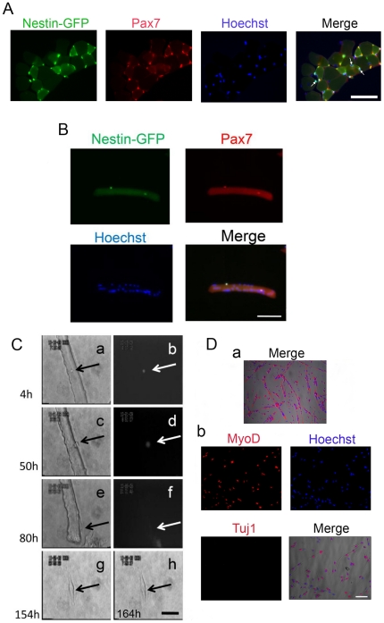
Figure 2. Nestin-GFP+ cells attached to myofiber (satellite cells) do not produce Tuj1+ cells with a neural phenotype.
A. A representative EDL muscle cross-section from a nestin-GFP transgenic mouse showing GFP expression and Pax7+ immunoreaction (n = 3 mice, 6 EDL muscles). Myofibers were counterstained with Hoechst. The merge image shows examples of GFP+/Pax- cells (arrows). Scale bar = 50 µm. B. Enzymatically dissociated single FDB muscle fiber showing 2 nestin-GFP+ satellite cells that immunoreact to Pax7 and overlap with Hoechst nuclear staining. Scale bar = 100 µm. C. FDB satellite cell time-lapse analysis. A nestin-GFP+ satellite cell attached to an FDB fiber (arrow) analyzed for more than 6 days. Snapshots of the complete record at 4, 50, 80, 154, and 162 h. Brightfield (a, c, e, g, h) and fluorescence (b, d, f). Fluorescence completely disappeared at 162 h in culture. Scale bar = 50 µm. Da. Four-month satellite cell culture from isolated EDL fibers. Myoblasts and myotubes are stained for desmin and Hoechst, which overlap with the brightfield image. b. The culture shown in Da is MyoD+ but Tuj1-. Scale bar for all pictures in D = 100 µm.

