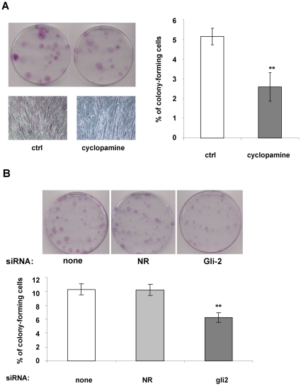
Figure 6. Inhibition of Hh signaling inhibits colony formation of hMADS cells.
Cells were plated at clonal density and maintained in medium supplemented with 10% FCS and FGF-2 (2.5 ng/ml) (Ctrl). One day after cell plating, 5 µM cyclopamine was added to the culture medium. (A): Photographs of colony formation of hMADS cells 3 weeks after treatment. A magnification of one colony is presented for each condition. (B): Photographs of colony formation of hMADS cells transfected with non relevant (NR) siRNA, Gli2 siRNA or only with hyperfect as a control (indicated “none” in the figure). Graphs represent the percentage of colony-forming cells in three independent experiments.

