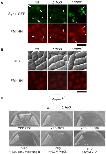
Figure 4. Rho3-deletion cells displayed phenotypes similar to those seen in Apm1-deletion cells.
(A) Wild-type (wt), Rho3-deletion (Δrho3), and Apm1-deletion cells (Δapm1) expressing chromosome-bone GFP-Syb1 cultured in YPD medium at 27°C were incubated with FM4-64 fluorescent dye for 5 min at 27°C to visualize Golgi/endosomes. GFP-Syb1 localization and FM4-64 fluorescence were examined under a fluorescence microscope. Bar 10 µm. (B) Wild-type (wt), Rho3-deletion (Δrho3), and Apm1-deletion cells (Δapm1) were cultured in YPD medium at 27°C. Cells were collected, labeled with FM4-64 fluorescent dye, resuspended in water, and examined by fluorescence microscopy. Photographs were taken after 60 min. Bar: 10 µm. (C) Wild-type (wt), Rho3-deletion (Δrho3), and Apm1-deletion cells (Δapm1) were streaked onto plates containing YPD or YPD plus 1.0 µg/mL micafungin, 0.5 µg/mL FK506, 0.3 M MgCl2, and 6 mM valproic acid, and were then incubated at 27°C for 4 d or at 36°C for 3 d.

