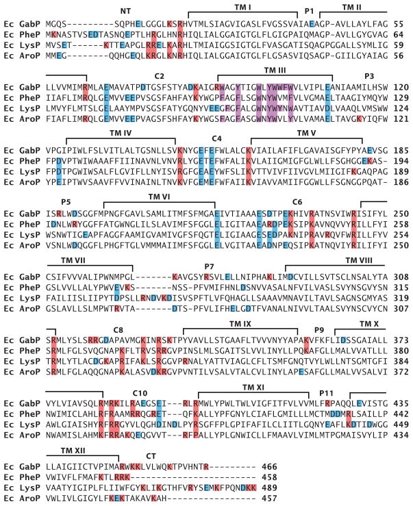
Figure 8.
Distribution of charged amino acids in homologous amino acid permeases. Sequence alignments shown for E. coli γ-aminobutyrate permease (GabP), phenylalanine (PheP), lysine (LysP), and aromatic (AroP) permeases. Positively or negatively charged amino acids are colored with red or blue, respectively, and aromatic residues in TM III are colored purple.

