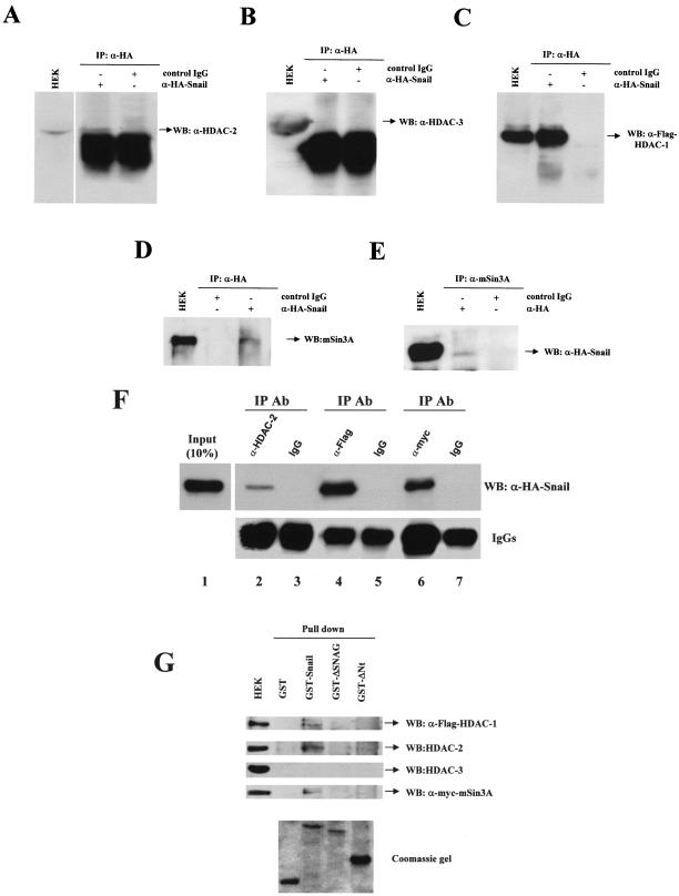
FIG. 4.
Snail associates with HDAC1/2 and the mSin3A corepressor through the SNAG domain. (A to D) HEK 293T cells were transiently transfected with Snail-HA and HDAC1-Flag constructs. Cell extracts were immunoprecipitated with anti-HA antibodies (α-HA) and control IgG, as indicated, and analyzed by Western blotting with anti-HDAC2 (α-HDAC-2), anti-HDAC3 (α-HDCA-3), anti-Flag (α-Flag), and anti-mSin3A (α-mSin3A) antibodies. (E) HEK 293T cells transiently transfected as above were immunoprecipitated with anti-mSin3A antibodies and analyzed by Western blotting with anti-HA. Cell extracts (HEK) were analyzed in parallel in all panels. (F) HEK 293T cells transiently transfected withSnail-HA, HDAC1-Flag, and mSin3A-myc constructs were immunoprecipitated with the indicated antibodies and analyzed by Western blotting with anti-HA antibodies (upper panel). Detection of IgG heavy chain is shown in the lower panel as an internal control. Input (10%) of whole cell extract was also analyzed in parallel. (G) Cell extracts obtained from HEK 293T cells transiently transfected as above were incubated with the indicated GST fusion proteins. The bound fractions from the glutathione-Sepharose beads and the input cell extract were analyzed by Western blotting with the indicated antibodies (upper panel). Analysis of the different recombinant GST fusion proteins used is shown in the lower panel. WB, Western blotting; IP, immunoprecipitation; Ab, antibody.

