
Figure 2.
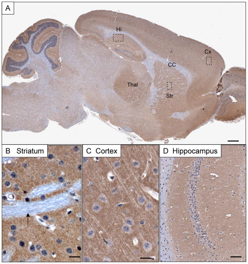
Overview of BAI1 immunoreactivity in the adult mouse brain. A) BAI1 expression detected by antibody h1570 is enriched in neuropil structures such as cortex (Cx), thalamus (Th), striatum (Str) and hippocampus (Hi), and largely absent from white matter structures such as the corpus callosum (CC). B) Striatal pencil fibers (small white matter tracts-arrow) lack BAI1 expression whereas surrounding neuropil shows finely punctate staining. C) Cortical pyramidal neurons show little somatodendritic cytoplasmic staining, but are surrounded by intense granular neuropil immunoreactivity. D) Hippocampal neurons show a neuropil pattern confined to the exent of the apical (left) and basal (right) dendritic fields of CA1. Scale bars: A=500 μm; B, C=20 μm; D=100 μm.