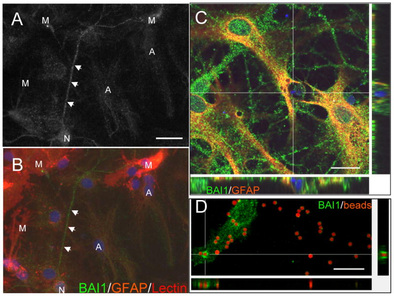
Figure 4.

(A,B) BAI1 is expressed by cultured neurons, astrocytes, and microglia. In general, neurons (N), recognized by their extensive neuritic networks, showed the strongest BAI1 immunoreactivity. Astrocytes (A), labeled lightly with GFAP-Cy3, also revealed significant punctate labeling. Microglia (M), labeled with the tomato lectin, showed variable and patchy BAI1 staining. (C) In older culture preparations in which numerous cells had undergone apoptosis, we frequently observed GFAP-positive astrocytes with punctate BAI1 labeling that had engulfed apoptotic nuclear and cytoplasmic debris. Confocal microscopy with z-sectioning showed the DAPI-positive nuclear debris to be within astrocyte cytoplasm. (D) BAI1-immunoreactive astrocyte membrane surrounds apoptotic targets (2 μm carboxylate-modified red fluorescent beads) added to cultured astrocytes for 2 hours. A single confocal slice shows that some beads are surrounded by a higher concentration of BAI1 staining (h1570) than surrounding plasma membrane (see bead at convergence of cross hairs). In addition, XZ and YZ orthogonal slices reveal BAI1 immunoreactivity surrounding a bead, consistent with its engulfment. Scale bars: A=25 μm, C=20 μm, D=15 μm.
