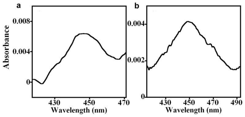
Figure 2. CO difference spectra of PDDA/PSS(/cyt P450 1A2/CPR+b5)6 films on aminosilane fused silica slides covalently cross-linked with poly(acrylic acid).

The cyt P450 heme in the PDDA/PSS(/cyt P450 1A2/CPR+b5)6 films were reduced directly with (a) 10 mM sodium dithionite, or (b) indirectly via CPR using 1 mM NADPH in anaerobic 50 mM phosphate buffer, pH 7.0, then purged 3 min with carbon monoxide (CO). Difference absorbance spectra with bands near 450 nm characteristic of native cyt P450s were acquired vs. similar PDDA/PSS(/cyt P450 1A2/CPR+b5)6 films that were reduced with sodium dithionite or NADPH, respectively, with no CO present. Denatured cyt P450s would give bands at 420 nm.
