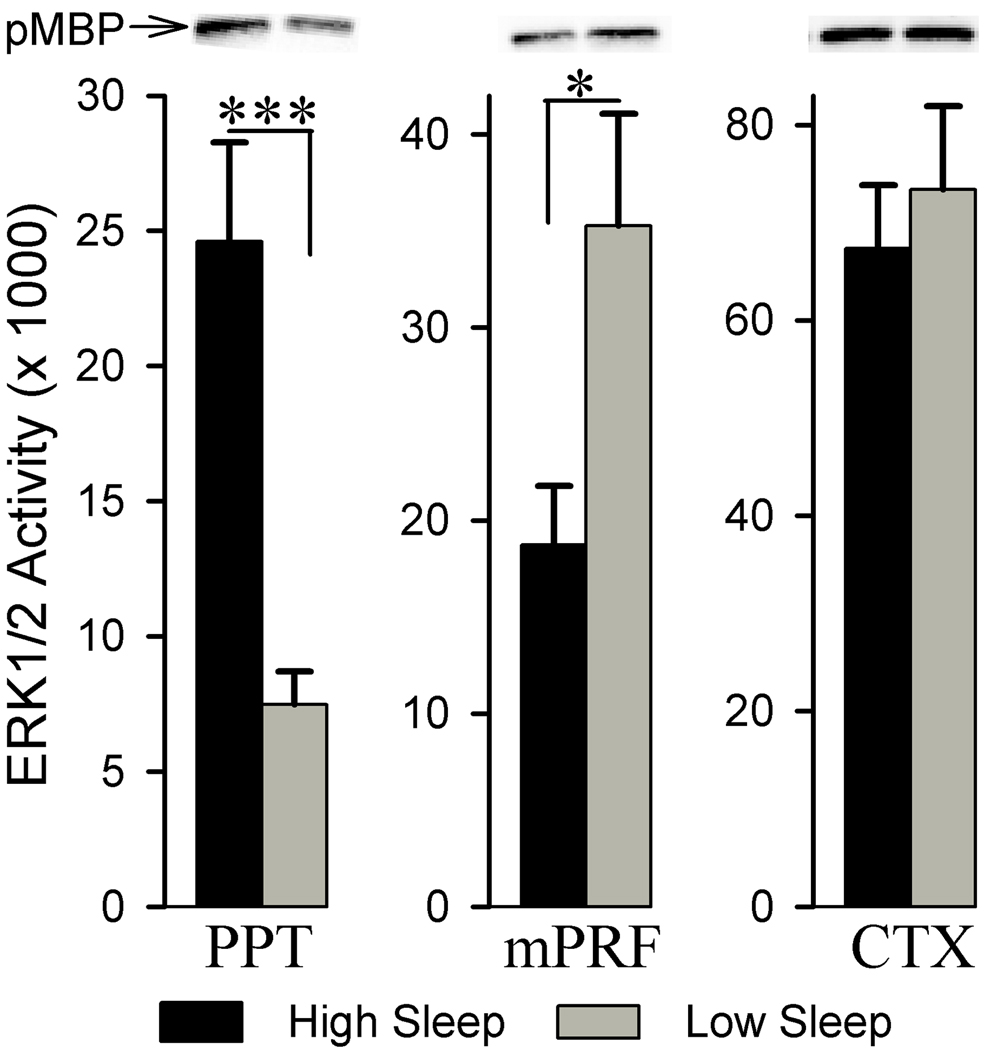
Figure 5.
Analysis of ERK1/2 activity in the pedunculopontine tegmentum (PPT), medial pontine reticular formation (mPRF), and cortex (CTX) of high and low sleep subjects. Densitometric measurements from western blots of phosphorylated myelin basic protein (pMBP) revealed that, compared to the low sleep group, in the high sleep group, ERK1/2 activity is higher (229.24% higher) within the PPT and lower within the mPRF (46.87% less). *** p<0.001, * p<0.05.

