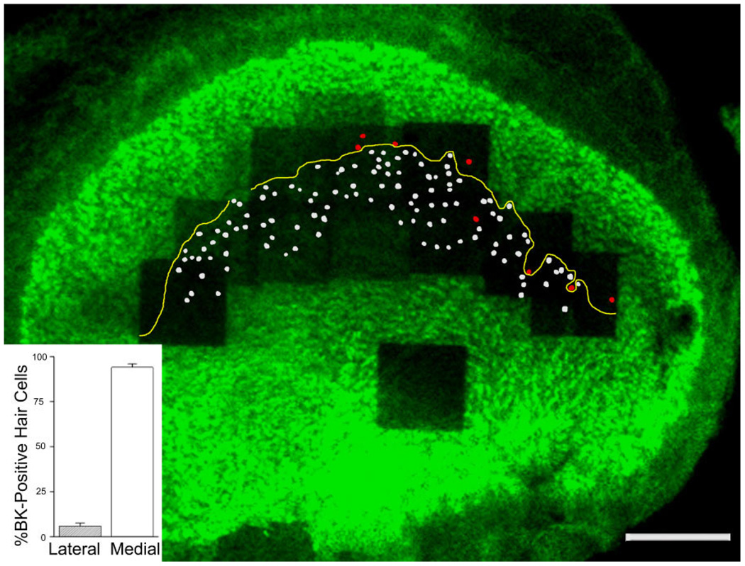
Figure 3.
Predominant localization of BK-positive hair cells on the medial side of the reversal line (yellow line). Hair cell tracings that have a lateral pointing MPV and thus are medial of the reversal line are indicated in white, whereas those that have a medial pointing MPV are colored in red. BK-positive hair cell quantification (inset) indicates that 94 ± 2% (n = 3 utricles from three animals) of all BK-positive hair cells in the utricle are medial of the reversal line. Note the single “orphan” hair cell with a medial-pointing MPV located medial to the reversal line. Scale bar = 100 µm).

