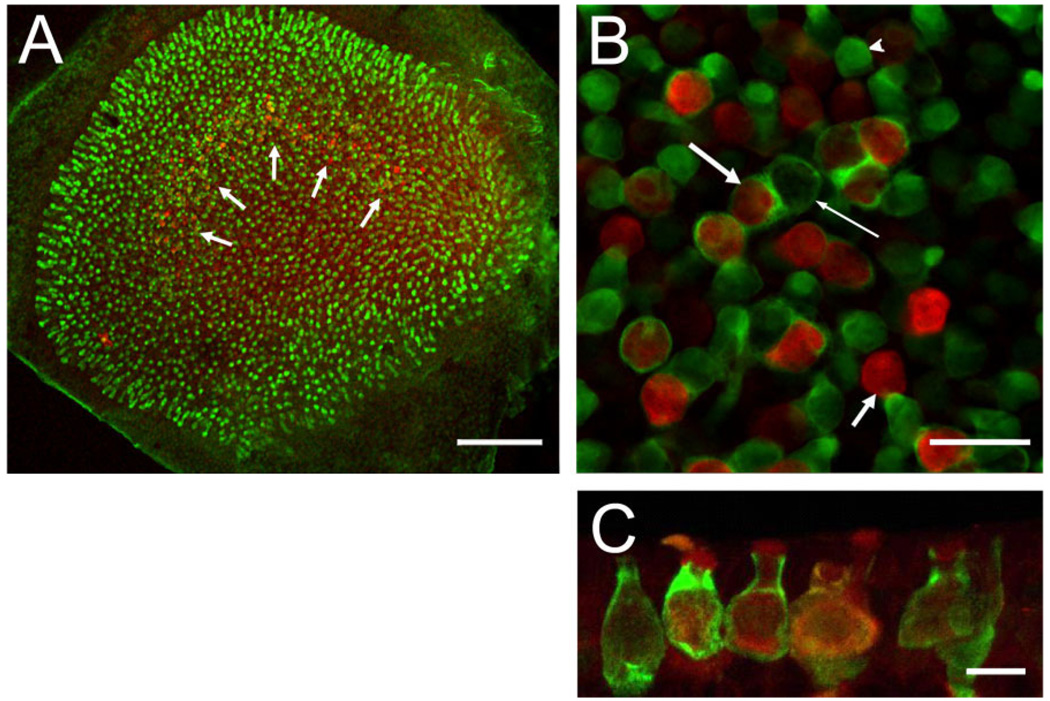
Figure 4.
Co-localization of BK channels (red) and calretinin (green). A: Whole-mount preparation viewed under low magnification. Many hair cells across the entire epithelium are calretinin positive (green), and a few calretinin-positive calyces can be discerned in the striolar region where a “band” of BK-positive hair cells is visible (white arrows). B: High-magnification view of striolar region. Note BK-positive hair cells surrounded (long, thick arrow) and not surrounded (short, thick arrow) by calretinin-positive calyces. Calretinin-positive calyces also surround BK-negative hair cells (thin arrow), and many calretinin-positive hair cells are visible (arrowhead). Of the BK-positive hair cells, 62 ± 4% (n = 3 utricles from 3 animals) are surrounded by a calretinin-positive calyx, characterizing them as type I. C: Maximum intensity projection of confocal images of cryostat section through the striolar region highlighting calyces and BK staining throughout hair cell (especially prominent at apical edge). Scale bar = 100 µm in A; 20 µm in B; 10 µm in C.

