Figure 5.
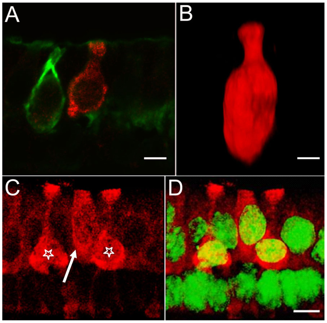
Diversity of BK-positive hair cells. Serially sectioned specimens immunolabeled with anti-BK (red) and anti-calretinin (green) antibodies illustrate the diversity of BK-positive hair cells. A: An example of a BK-positive type I hair cell not encapsulated by a calretinin-positive calyx. The BK-positive hair cell exhibits the flask-shaped morphology indicative of a type I hair cell. Note the absence of BK-labeling in the adjacent hair cell enveloped by the calretinin-positive calyx. B: Three-dimensional volume reconstruction of a BK-positive hair cell from a different specimen. The classical type I morphology of this hair cell can be clearly appreciated. C: A putative BK-positive type II cell (arrow) next to two BK-positive type I cells (stars). D: Same image as in C, but with superimposed nuclear staining (NeuroTrace, green). Note the apical localization of the nucleus for the putative type II hair cell. Scale bar = 5 µm.

