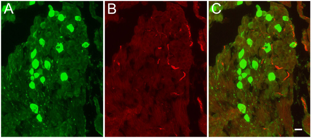
Figure 6.
Absence of BK immunoreactivity in Scarpa’s ganglion. Cryosections through Scarpa’s ganglion were double stained for (A) calretinin (green, left) and (B) BK (red, middle). C: Superposition of images shown in A and B. Note the absence of BK-positive cell bodies even at high exposure times. Scale bar = 10 µm.

