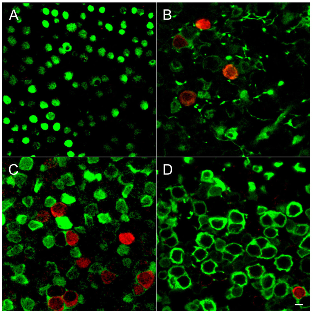
Figure 7.
Age-dependent expression of BK channels in the utricle. Whole-mount preparations of utricles were immunolabeled for BK (red) and calretinin (green). Single confocal sections taken from the striolar region are shown. A: At P6 calretinin-positive calyces are not well developed, making it difficult to identify the striolar region. However, no BK-positive hair cells were observed in any part of the epithelium. B: By P14 BK-positive hair cells are surrounded by calretinin-positive calyces. We rarely observed four BK-positive hair cells in a single field of view. C: By P21 many BK-positive hair cells engulfed in calretinin-positive calyces are present. D: By 2 months of age (P62) only a few BK-positive hair cells are evident. Scale bar = 5 µm in A–D.

