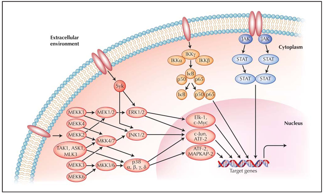
Figure 1.
Schematic of intracellular signaling cascades. Cellular exposure to cytokines, chemokines, growth factors, pathogen-associated molecular patterns or antigens, endogenous danger signals, or stressors such as ultraviolet rays or absence of growth factors results in receptor ligation. Subsequent initiation of signaling cascades leads to altered expression patterns of genes involved in inflammation, degradation of extracellular matrix, apoptosis, and other cellular processes important in mounting an appropriate response to the stimuli. ASK—apoptosis signal-regulating kinase; ATF—activating transcription factor; ERK—extracellular signal-regulated kinase; IκB—inhibitor of nuclear factor κ-light-chain-enhancer of activated B cells (NF-κB); IKK—IκB kinase; JAK—Janus tyrosine kinase; JNK—c-Jun N-terminal kinase; MAP-KAP— mitogen-activated protein kinase (MAPK) activated protein; MEK—MAPK/ERK kinase; MEKK—MAPK kinase kinase/MEK kinase; MLK—mixed lineage kinase; MKK—MAPK kinase; STAT—signal transducer and activator of transcription; Syk—spleen tyrosine kinase; TAK—transforming growth factor-β–associated kinase.

