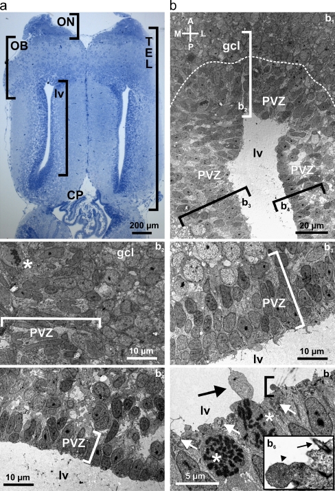
Fig. 1.
Ultrastructural analysis of the anterior telencephalon. a Horizontal slice of the telencephalon of larval X. laevis stained with Methylenblue-Azur II. b Transmission electron microscopy of the anterior part of the lateral telencephalic ventricles with its PVZ and the posterior part of the granule cell layer of the OB (b 1). Various electron micrographs were assembled into a photo montage to obtain this overview. The dotted line indicates the approximate border between the PVZ and the granule cell layer of the OB. The brackets indicate the approximate location of the areas shown at higher magnification in b2 through b4. Anterior part of the PVZ and adjacent granule cell layer of the OB (b 2). Cell divisions are frequent in the PVZ. Note the cell with decomposed nuclear envelope and condensed DNA in form of chromosomes (asterisk). Medial part of the PVZ (b 3) and lateral part of the PVZ (b 4). The brackets in b 2 through b 4 include the cell layers of the anterior, medial, and lateral PVZ, respectively. b5 shows a higher magnification of cells in direct contact with the lumen of the lateral ventricles. Two cells are in mitosis (asterisks). The white arrows indicate accumulations of mitochondria very close to the ventricular lumen. The black arrow shows a large cellular protrusion into the ventricle. The area included in the bracket is shown at higher magnification in b6. Note the multivesicular protrusion into the ventricle (arrowhead) and the single cilium extending into the cerebrospinal fluid (arrow). Abbreviations: ON olfactory nerve, OB olfactory bulb, PVZ periventricular zone, TEL telencephalon, CP choroid plexus, lv lateral ventricle, gcl granule cell layer

