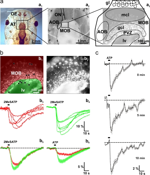
Fig. 3.
Nucleotide-induced [Ca2+]i increases in cells of the anterior telencephalon. a Overview of a head of larval X. laevis (a 1). The black rectangle indicates the location of the anterior telencephalon. An acute slice preparation of the telencephalon is shown in a 2. The black rectangle indicates the approximate location of the Fura-2/AM stained slice shown in a3 (brightened fluorescence image acquired at rest). b Fura-2/AM stained slice preparation (the same as shown in a3) with the area of the OB shaded in red, and the PVZ shaded in green (b 1). Pixel correlation map of the same slice (see “Materials and methods” section for details) obtained upon application of 2MeSATP (5 μM; b2). The ventricle appears to respond as a whole. This results from responses of ependymal cells from the floor of the lateral ventricle. The 2MeSATP-induced [Ca2+]i transients of six individual cells of the OB (see black dots in the red-shaded area in b1) and the PVZ (see black dots in the green shaded area in b1) are shown in b3 and b4, respectively. The 2MeSATP-induced mean [Ca2+]i transients (±SEM) of all clearly identifiable responsive cells of the OB (red traces; n = 138) and the cells of the PVZ (green traces; n = 32) of this slice are shown in b5. Application of ATP (50 μM) to the same slice preparation induced comparable [Ca2+]i transients in the same cells as above (b 6). (c) Mean [Ca2+]i transients of responding OB and PVZ cells (n = 120) remain stable after repeated applications of ATP (100 μM; interstimulus interval: 5 min). Abbreviations: OE olfactory epithelium, AT anterior telencephalon, ON olfactory nerve, MOB main olfactory bulb, AOB accessory olfactory bulb, PVZ periventricular zone, gl glomerular layer/glomeruli, mcl mitral cell layer, gcl granule cell layer, lv lateral ventricle

