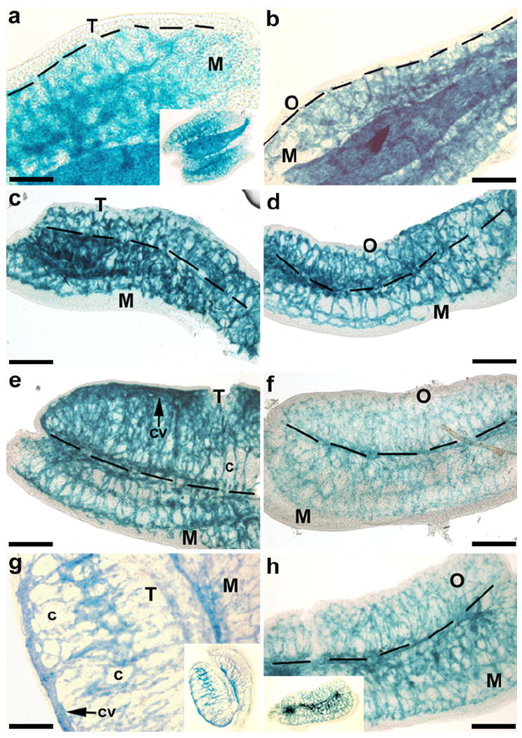
Fig. 1.

Whole-mount images of gonads from embryonic day 11-13 (E11-13) kinase insert domain protein receptor (KDR)-LacZ mice. In the genital ridges from E11 male (a) and female (b) mice, KDR-LacZ staining was located predominately within the vasculature of the mesonephros (M). Staining patterns and organ sizes were similar between testes (T) and ovaries (O) of E12 mice (c, d). By E12.5, the testis (e) had surpassed the ovary (f) in size, and the vascular pattern began to become distinct in the testis with the formation of a coelomic vessel (cv) and vasculature surrounding the seminiferous cords (c). At E13, the testis (g) was larger and more rounded in shape with more defined vasculature around the seminiferous cords, while the vasculature within the ovary and mesonephros of the female (h) was much more densely organized than in the male. Insets Low-power views. Bars 200 μm
