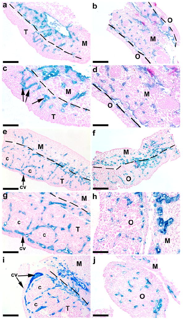
Fig. 2.

Expression of KDR-LacZ in the mesonephroi and gonads from E11.5-13.5 mice. In E11.5 testes (a, c), small populations of KDR-expressing cells (arrows) appeared in the testis (T), in close proximity to the mesonephros (M). By E12.5 (e, g) and E13.5 (i, cross section), the coelomic vessel (cv) had developed in the testis, and KDR-expressing cells surrounded the seminiferous cords (c). In E11.5 females (b, d), KDR-LacZ staining was located predominately within the vasculature of the mesonephros. Vascular development was evident at E12.5 (f) in the ovary (O). At E13.5 (h, j, cross sections), KDR-LacZ staining was still present throughout the ovary. Bars 100 μm (a-b, e-f), 50 μm (c-d, g-j)
