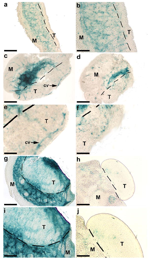
Fig. 3.

Effects on KDR-LacZ expression in E11 mouse organ cultures after treatment with a vascular endothelial growth factor A (VEGFA) receptor tyrosine kinase antagonist (VEGFR-TKI). Immediately before culture (a, b), KDR-LacZ staining was located predominantly in the mesonephros (M) with only a few KDR-expressing cells in the testis (T). After 1 day in culture (D1), endothelial cells had established vascular patterns, including a distinct coelomic vessel (cv), within the control testis (c, e). Treated testes contained KDR-expressing cells on D1, but in fewer numbers and without distinct patterns (d, f). By D3 of culture, KDR-LacZ was still present in control testes (g, i), whereas treated testes (h, j) had little apparent staining. Bars 200 μm (a, c-d), 100 μm (b, e-f, i-j), 50 μm (g-h)
