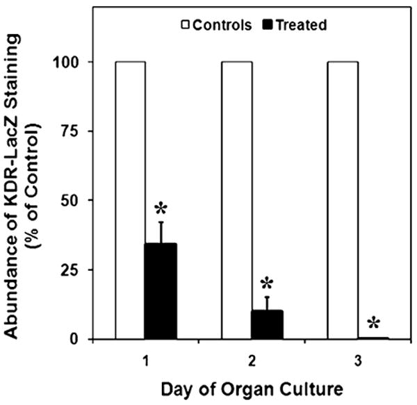
Fig. 4.

Effects of VEGFR-TKI treatment on KDR-LacZ staining in cultured E11 testes, expressed as a percentage of control organs. After days 1, 2, and 3 of culture, β-galactosidase staining was reduced by 66%, 90%, and 99%, respectively, in treated testes. The mean areas of staining (±SEM) are presented. *Statistically significant difference compared with the means of controls at P<0.002
