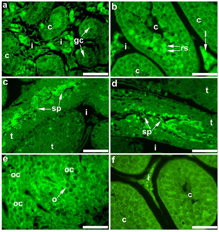
Fig. 8.

Immunohistochemical staining for β-galactosidase in embryonic and postnatal mouse gonads. In E17 testes (a), staining was detectable around germ cells (gc) within the seminiferous cords (c), and intense staining was present in the interstitium (i). In the postnatal testis, β-galactosidase was localized to Leydig cells (l) in the interstitium and round spermatids (rs) within cords at P20 (b) and in sperm (sp) within seminiferous tubules (t) at P60 (c, d). Immunohistochemistry for β-galactosidase in the E17 ovary (e) revealed positive staining around oocytes within oocyte cysts (oc). Gonadal sections with no primary antibody served as negative controls; a P20 testis section is shown (f). Bars 100 μm (c), 50 μm (a, b, d-f)
