FIG. 6.
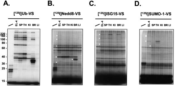
UBL-VSs exhibit differential labeling patterns in mouse tissue extracts. Lysates from single-cell suspensions of the indicated mouse tissues were prepared and incubated for 1 h at 37°C with radiolabeled UBL-VSs. Per reaction, 5 × 10⁵ cpm of 125I-UBL-VS and 20 μg of EL-4 or tissue lysate were used. The reactions were terminated as described for Fig. 2. Polypeptides were resolved by SDS-10% PAGE and visualized by autoradiography. First lane in each panel, no lysate added (UBL-VS probe only). SP, spleen; TH, thymus; KI, kidney; BR, brain; LI, liver. Triangles mark the positions of observed UBL-VS-protein adducts. The radiolabeled UBL-VS probes used were125I-Ub-VS (A), 125I-Nedd8-VS (B), 125I-UCRP-VS (C), and 125I-SUMO-1-VS (D). The positions of molecular mass markers (in kilodaltons) are indicated on the left.

