Fig. 1.
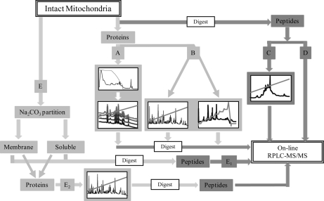
Schematic representation of multidimensional fractionation of mitochondria. Mitochondria were subjected to five orthogonal approaches. Following initial preparation of intact mitochondria, a small subset was further fractionated on the basis of topographical location (E). For protein-centric analysis (light gray), proteins were solublized and subject to either 2-DLC (A; HPCF and RP-HPLC) or 1-DLC (B; RP-HPLC alone). 1-DLC was performed using both linear and sawtooth RP gradients. Each resulting fraction was then digest and subject to RPLC-MS/MS. Peptide-centric analysis (dark gray) was performed on mitochondrial tryptic peptides. Samples were fractionated by 2-DLC (C; SCX and on-line RPLC-MS/MS) and 1-DLC (D; on-line RPLC-MS/MS alone). Subfractionated mitochondria (E) whereby membrane-associated and soluble partitions were created, were separated by either peptide-centric 1-DLC (E1) or protein-centric 1-DLC (E2). Protein-centric 2-DLC of intact mitochondria (A) and protein-centric 1-DLC of fractionated mitochondria (E2) are the strategies that resulted in three dimensions of fractionation.

