Figure 3.
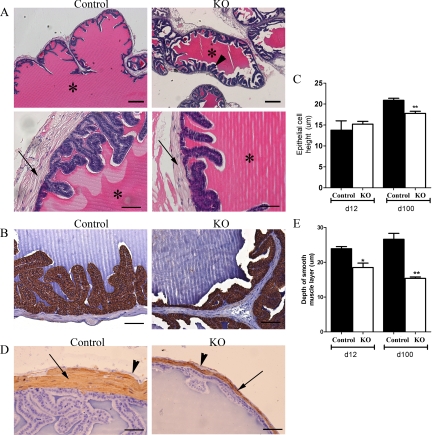
Histological analysis of PTM-ARKO seminal vesicles. A, Hematoxylin and eosin staining of d 100 PTM-ARKO and control SVs composed of epithelia surrounded by a stromal compartment (arrow); this epithelium appeared more folded in PTM-ARKO SVs than control (arrowhead). Note the dense eosinophilic seminal secretions in the lumen of both the PTM-ARKO and control SV (*). Scale bars, 400 and 50 μm, respectively. B, Immunohistochemistry for cytokeratin (brown), highlighting the epithelial cells in d 100 PTM-ARKO and control SVs. C, Note that epithelial cell height is significantly reduced in PTM-ARKOs at d 100 but not d 12, compared with age-matched controls. Scale bars, 50 μm. D, SMA immunostaining (brown) identifying the smooth muscle cell layer (arrow) directly surrounding the epithelium in d 100 PTM-ARKO and control SVs; note the presence of an outer SMA-negative layer (arrowhead). E, The smooth muscle layer is narrower in the PTM-ARKO than the control, as confirmed quantitatively. Values are means ± sem (n = 3 mice). *, P < 0.05, **, P < 0.01, compared with age-matched controls. Scale bars, 50 μm.

