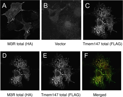
Fig. 6.
Effect of Tmem147 expression on the subcellular distribution of the M3R in cotransfected COS-7 Cells. The subcellular localization of the M3R-HA receptor, either expressed alone or coexpressed with Tmem147-FLAG was studied via confocal microscopy using transfected COS-7 cells. After fixation, cells were permeabilized with 0.2% saponin. A, transfection with the M3R-HA construct alone. Cells were stained with a monoclonal anti-HA antibody followed by incubation with a secondary antibody. Note the intense staining of the M3R-HA protein on the cell surface. B, transfection with vector DNA (pcDNA3.1; control). Cells were treated with the anti-HA antibody to reveal background staining. C, transfection with the Tmem147-FLAG construct alone. Tmem147-FLAG was visualized by using a polyclonal anti-FLAG antibody followed by incubation with a secondary antibody. Note that Tmem147 protein was retained intracellularly (in the ER; see Fig. 5). D to F, cotransfection of COS-7 cells with the M3R-HA and Tmem147-FLAG constructs. Cells were stained with the indicated antibodies. Note that cell surface expression of the M3R-HA receptor was barely visible in the presence of coexpressed Tmem147-FLAG (D) and that the two proteins were colocalized in the ER (D–F). For experimental details, see Materials and Methods.

