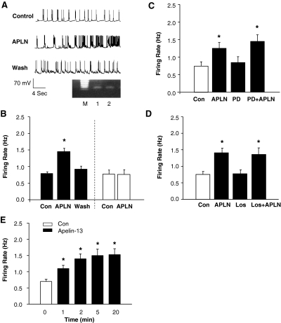
Fig. 2.
Apelin-13 exerts a positive chronotropic action via an AT1 receptor- and AT2 receptor-independent mechanism. A, representative recordings showing spontaneous neuronal action potentials recorded under the following conditions: superfusion of control solution (PBS), superfusion of apelin-13 (APLN, 100 nM), and followed by washing with superfusate solution. Action potentials were recorded from an APJ receptor-positive neuron identified by single-cell RT-PCR. Ethidium bromide-stained gels, seen in A, show the PCR DNA products that correspond to APJ receptor mRNA obtained from the same neuron after electrophysiological recordings (lane 2). The RNA from the whole dish of neurons was used as positive control (lane 1). M, marker. B, bar graph summarizing the effect of apelin-13 on the neuronal firing rate recorded from APJ receptor-positive neurons (filled bars) or APJ receptor-negative neurons (open bars) identified by RT-PCR. Data are means ± S.E. (n = 7, 6). *, P < 0.01 compared with Con. C, effect of apelin-13 on neuronal firing rate under the following sequential treatment conditions: superfusion with control (Con) solution (PBS), superfusion with apelin-13 (APLN, 100 nM), wash with superfusate solution, superfusion with PD123319 (PD, 1 μM), and combined application of apelin-13 and PD123319 (PD+APLN). Data are mean ± S.E. from seven neurons. *, P < 0.01 compared with Con. D, bar graphs of the firing rate recorded in each treatment condition as described in C, with the exception of PD123319, which was replaced with losartan (Los, 1 μM). Data are mean ± S.E. from seven neurons. *, P < 0.01 versus respective control treatment. E, time-dependent response of neuronal firing to apelin-13. The neuronal firing was recorded in neurons treated with apelin-13 (100 nM) for the time periods indicated in the graph. Results are mean ± S.E.; n = 6. *, P < 0.05 compared with Con.

