
Figure 1.
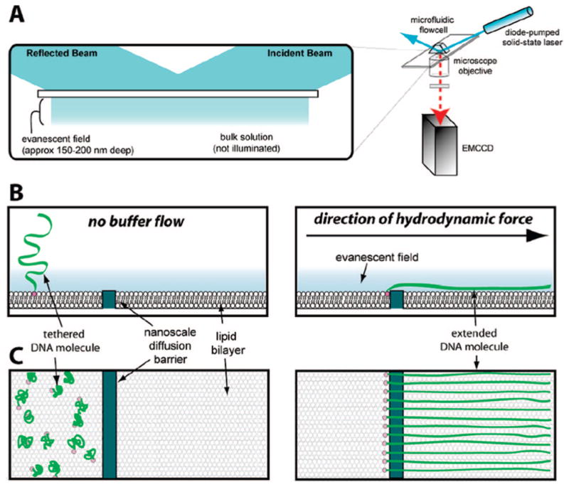
Conceptual diagram of lipid tethered DNA molecules aligned at a diffusion barrier. Panel (A) shows a diagram of the total internal reflection fluorescence microscope (TIRFM) used to image single molecules of DNA. For imaging by TIRFM the long DNA molecules (48 kb) used in these studies must be extended parallel to the surface of the sample chamber in order to remain confined within the evanescent field. Panels (B) and (C) depict a cartoon illustration of the bilayer on the surface of a fused silica slide along with a barrier and the response of tethered DNA molecules to the application of a hydrodynamic force. The upper and lower panels in (B) and (C) depict views from the side and above, respectively. In the absence of buffer flow (B) the DNA molecules are tethered to the surface, but are not confined within the evanescent field, nor are they aligned at the barrier. As depicted in (C), when flow is applied, the DNA molecules are dragged through the bilayer until they encounter the diffusion barrier, at which point they will align with respect to one another and form a curtain of DNA molecules. Please note that these drawings are not to scale.