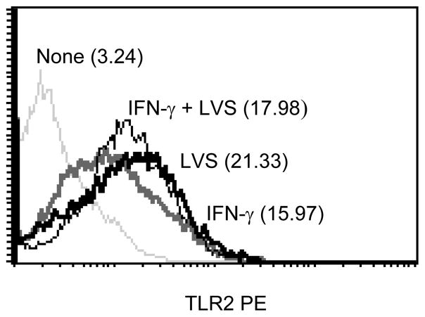
Figure 10.
The role of IFN-γ on F. tularensis-induced TLR2 expression on macrophages. Macrophages from C57BL/6 wt mice were cultured with IFN-γ (200 U/ml) either alone or in combination with F. tularensis LVS (MOI = 10) for 24 h. The cells were stained with PE-conjugated anti-mouse TLR2 antibody and surface expression of TLR2 was determined by flow cytometric analysis. The values in the histograms are the MFI. The results are representative of three separate experiments.

