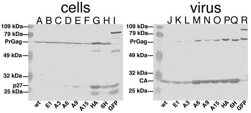
Figure 2. Cell and viral proteins.
Cell lysate (A-I) and virus (J-R) samples from 293T cells transfected with different pXM-GPE gag-pol-env expression constructs were collected 72 h post-transfection. Proteins in samples were separated by SDS-PAGE, and viral proteins were detected by immunoblotting with a primary anti-p12 antibody (A-I) or a primary anti-CA antibody (J-R). Viral variants from which the samples are derived are indicated on the bottom of each panel. At the left hand side of each panel are prestained size standards, and the sizes of the markers are as indicated. Also indicated are the migration positions of WT PrGag, viral CA, and cellular p27 (MA plus p12) proteins.

