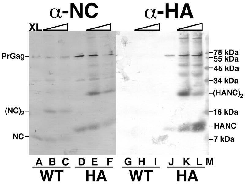
Figure 4. Crosslink analysis of NC proteins.
WT (A-C, G-I) and HA (D-F, J-L) virus samples were mock-treated (A, D, G, J) or crosslinked with 0.1 mM (B, E, H, K) or 1 mM (C, F, I, L) BMH. After crosslinking, protein samples were separated by SDS-PAGE on a 16% acrylamide gel, and proteins were detected by immunoblotting with primary anti-NC antibodies (A-F) or a primary anti-HA antibody (G-L). Marker proteins were separated in lane M, and their sizes are indicated on the right hand side. Also indicated are the migration positions of the PrGag proteins, the WT monomer (NC) and dimer ([NC]2) proteins, and the HA nucleocapsid monomer (HANC) and dimer ([HANC]2) proteins.

