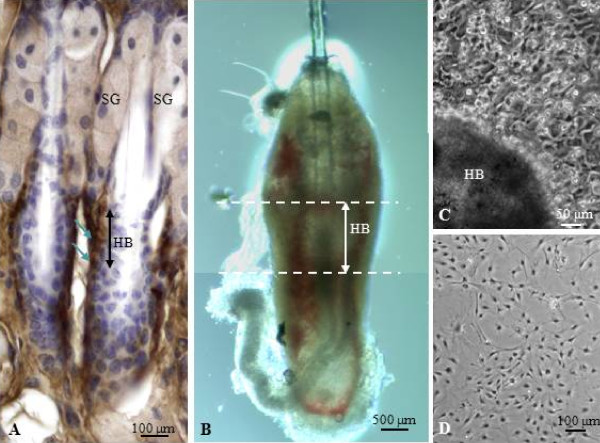
Figure 1.
Establishment of primary hair bulge cell culture. (A) Immunohistological staining revealed a large number of CD34+ cells (arrows) present in the vicinity of the hair bulge (HB) inferior to sebaceous glands (SG). (B) Showing an isolated hair follicle from mouse vibrissal hair. The hair bulge was dissected (extent indicated by the dotted lines) from the follicle and used for explant culture. (C) Showing cells that have migrated out from the bulge explant to form colonies of cells all around the explant. (D) Showing the appearance of HBPCs following purification with anti-CD34 conjugated magnetic beads.

