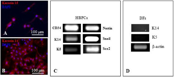
Figure 2.
Characterization of CD34+ HBPCs. (A-B) Immunofluorescent staining showed HBPCs specifically expressed established HBPCs surface markers Keratin 14 (K14) and 15 (K15). (C) Semi-quantitative RT-PCR analysis confirmed that the HBPCs expressed K14. In addition, HBPCs also expressed K5 and Snail and Sox2. (D) In contrast, dermal fibroblasts (DFs) isolated from adjacent to the hair bulge did not express K14 and K5. β-actin served as an internal control.

