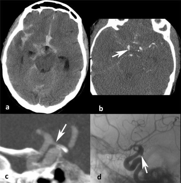
Figure 1.

Missed aneurysm. Aneurysm missed by each neuroradiologist on the CT Angiography (CTA). a Non-contrast CT displaying the aneurismal SAH pattern. b CTA showing the posterior communicating aneurysm (arrow). c 3D reconstruction of the CTA showing the aneurysm filled with contrast material, appearing to be continuous with the sphenoid bone (arrow). d Angiogram of the same 3 mm aneurysm on the posterior communicating artery (arrow)
