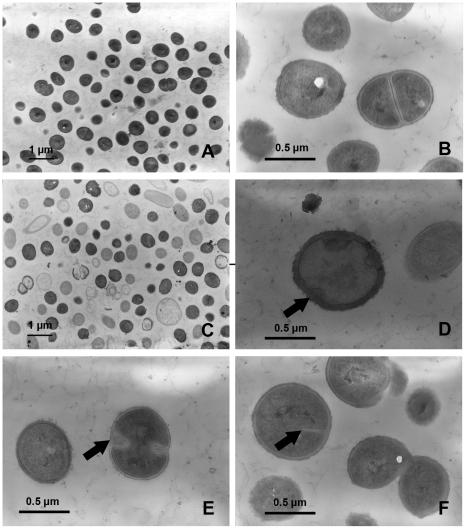
Figure 3. Transmission electron microscopy demonstrating the effects of rhodomyrtone on methicillin-resistant S. aureus NPRC 001R morphology and ultrastructure.
The bacteria were incubated in CAMHB for 18 h, media containing 0.174 µg/ml of rhodomyrtone (C, D, E, and F) and untreated control cultures (A and B). Scale bars = 1 µm (A and C) and 0.5 µm (B, D, E, and F), respectively.

