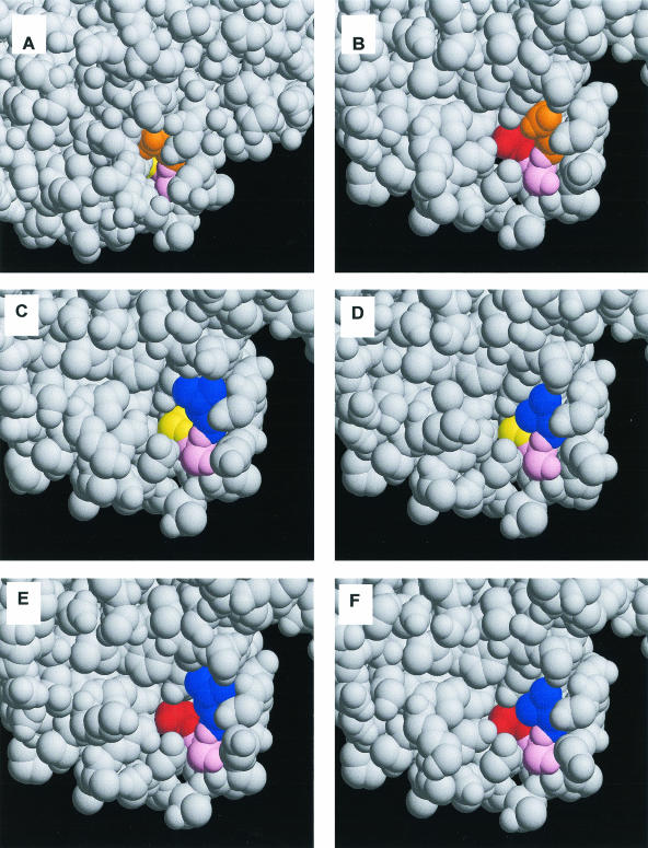
FIG. 2.
Modeling of point mutations into the receptor binding site of H5 HA. (A) Wild type; (B) G228S mutant; (C) Q226L mutant; (D) Q226V mutant; (E) LSS double mutant; (F) VSS double mutant. Native residues 226Q, 227S, and 228G are shown in orange, pink, and yellow, respectively. Mutations to a hydrogen bonding residue (serine) are shown in red, and mutations to a hydrophobic residue (leucine-valine) are shown in blue.

