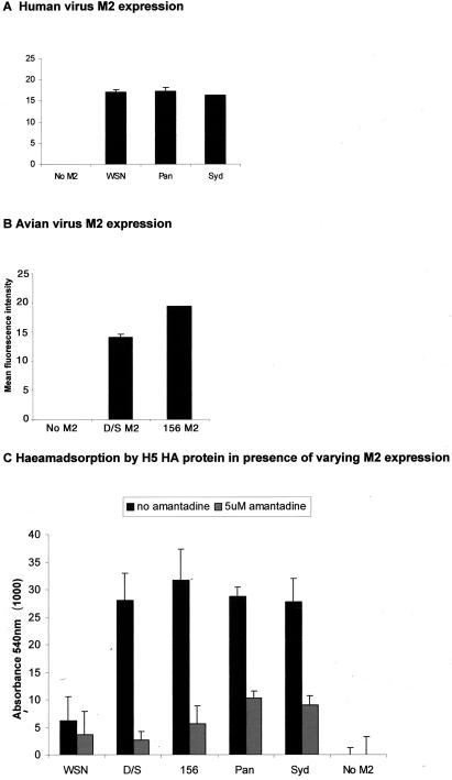
FIG. 3.
(A and B) Human and avian virus M2 protein expression. Forty-eight hours following transfection of 0.2 μg of plasmid DNA, cells were incubated with primary antibodies against M2. A mouse monoclonal antibody (14C2) was used to detect the human M2 proteins (A), and a rabbit polyclonal antiserum raised against a swine influenza virus M2, kindly provided by P. Heinen, was used to detect the avian M2 proteins (B). Fluorescein isothiocyanate-conjugated secondary antibody was used, and the mean fluorescence intensity of each sample was measured by fluorescence-activated cell sorter analysis. (C) Representation of the ability of different M2 proteins to support hemadsorption by H5 HA. Vero cells were transfected with 1 μg of plasmid directing the expression of HK156 HA and 0.2 μg of plasmid expressing different avian or human viral M2 proteins and incubated either with (grey bars) or without (black bars) 5 μM amantadine. Forty-eight hours posttransfection, cells were treated with 5.5 mU of bacterial neuraminidase (V. cholerae sialidase) per ml, and a 0.5% suspension of horse erythrocytes was added. After 1 h of incubation at room temperature, cells were washed and lysed for absorbance measurement at 540 nm. Cells infected with FPV T7 but untransfected were processed in the same way, and the mean absorbance from these wells was subtracted as background. All transfections were performed in triplicate, and mean absorbance values with standard errors are shown. D/S, A/Duck/Singapore/3/97; 156, A/HK/156/97; Pan, A/Panama/99; Syd, A/Sydney/5/97.

