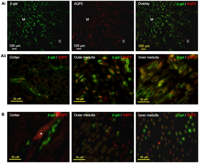
Figure 7. Localization of β-galactosidase (β-gal) to principal cells in collecting ducts.
Ai. In kidneys of 2-week-old mice, immunostaining pattern of β-gal (green) closely resembled that of aquaporin 2 (AQP2, red). Right panel showed co-localization of the two signals to the same tubules. Original magnification was 100×. Aii. Merged images: Principal cells in the cortex, outer medulla and inner medulla appeared yellow-orange when AQP2 signal co-localized with β-gal signal. Original magnification was 400×. B. β-gal (green) signal was also observed in principal cells that stained positive for AQP2 (red) in the collecting ducts in cortex, outer medulla and inner medulla of 8-week-old mice. Asterisk indicates non-specific staining, which was also observed on sections incubated with non-immune IgGs in place of primary antibodies (data not shown). Original magnification was 400×.

