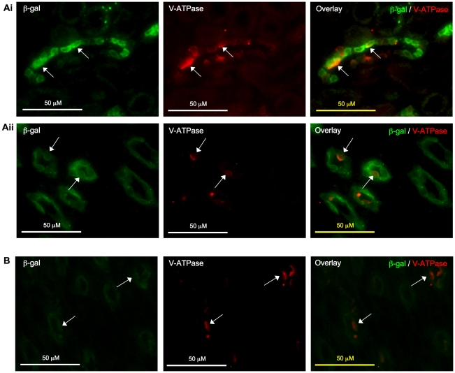
Figure 8. Localization of β-galactosidase (β-gal) to intercalated cells in collecting ducts.
β-gal signal (green) was detected in intercalated cells of collecting ducts (arrow) that stained positive for vacuolar H+-ATPase B1 (V-ATPase, red) in kidney cortex (Ai) and medulla (Aii) of 2-week-old mice. In 8-week-old mice, β-gal signal (green) was less apparent in the cortex but was noted in the medulla (arrow) (B). Original magnification was 400×. No specific signal was detected on sections incubated with non-immune IgGs in place of primary antibodies (data not shown).

