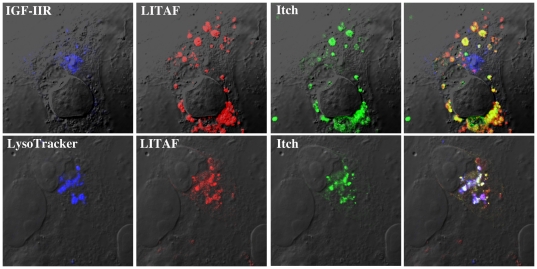
Figure 3. LITAF changes the cellular localization of Itch.
FLAG-LITAF was transiently co-transfected into BGMK cells with GFP-Itch. Sixteen hours post-transfection, cells were probed with LysoTracker (blue) and fixed. LITAF was detected using anti-FLAG antibodies (red) while the trans-Golgi network was identified using anti-IGF-IIR antibodies (blue). GFP-Itch is shown in green. Cells were visualized using DIC.

