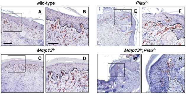
Figure 4. Angiogenesis in skin wounds in Mmp13;Plau double-deficient mice.
Angiogenesis in wounds removed 14 days after incision is visualized by immunostaining of endothelial cells with antimouse CD34 in wild-type (A+B), Mmp13-deficient (C+D), Plau-deficient (E+F) and Mmp13;Plau double-deficient (G+H) mice. The box insets in (A, C, E+G) indicate the magnified views shown in (B, D, F+H). In (F+H) the arrows mark where the vessels protrude into the epidermal layer of the wounds. The scale bar in (A) = 0.2 mm and is representative also for (C, E+G). The scale bar in (B) = 0.1 mm and is representative also for (D, F+H).

