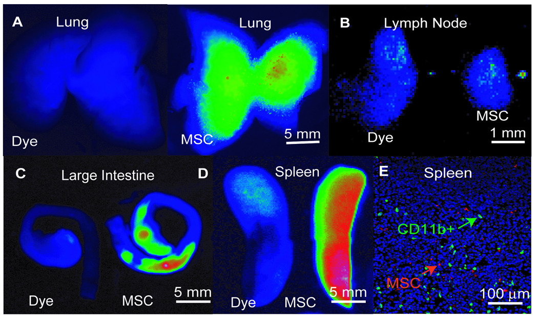
Figure 5. Near Infrared Tracking of MSC Transplants.

MSCs were labeled with VT680 and infused intravenously at the time of TNBS administration. Control images, labeled as ‘dye’, are injections of VT680 dye alone. Representative images from the (A) lungs, (B) mesenteric lymph nodes (MLN), (C) large intestine, and (D) spleens of mice injected with the dye alone or labeled MSCs one day post-infusion. Results of two independent imaging studies of N=3 per group. (E) Immunofluorescent micrographs of splenic tissue harvested from mice infused with VT680-labeled MSCs (red) and co-stained for CD11b (green), and DAPI (blue).
