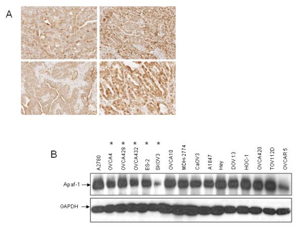
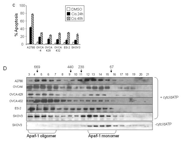
Figure 1. Apaf-1 expression is not predictive of response to cisplatin.


A. Tissue microarrays containing cores from 87 epithelial ovarian tumors were analyzed for Apaf-1 expression with immunohistochemistry. Representative samples shown. B. Immunoblot analysis of cytosolic Apaf-1 expression in ovarian cancer cell lines. The level of Apaf-1 in a panel of ovarian carcinoma cell lines was determined by immunoblotting.
Chemoresistant cell lines are indicated *. C. Representative ovarian carcinoma cell lines were treated with DMSO or cisplatin (5 μg/ml) for 24 or 48 hours. The percent of apoptotic cells was determined by propidium iodide staining and subG0 analysis. Values are expressed as mean±SD (n= 3). D. A. Cytosolic extracts from chemosensitive (A2780) and chemoresistant (OVCA 4, OVCA 429, OVCA 432, ES-2, and SKOV3) cell lines were incubated for 30 minutes in the presence or absence of cytochrome c and dATP. Extracts were fractionated on a Superdex 200 HR column. Equal amounts (50 μl) of each fraction were resolved by SDS PAGE and Apaf-1 was detected by immunoblotting. The elution profiles and sizes of selected standards are indicated.
