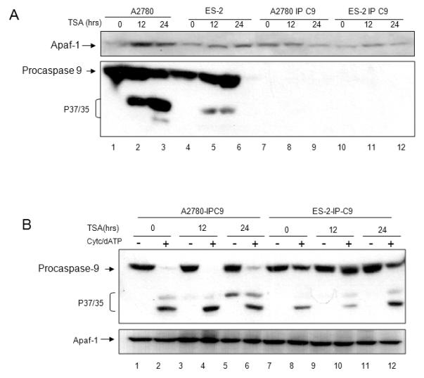
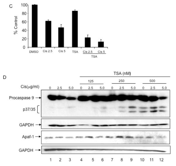
Figure 6. TSA treatment increases Apaf-1 activity.


A. A2780 and ES-2 cells were treated with TSA (0.5 μM) for 0, 12, and 24 hours. Cellular lysates were immunodepleted of caspase-9 (A2780-IP-C9, ES-2-IP-C9). Apaf-1 and caspase-9 expression levels were determined by immunoblotting. B. A2780-IP-C9 and ES-2-IP-C9 lysates were normalized for Apaf-1 expression (bottom panel). Lysates were incubated with in vitro translated pro-caspase-9 in the presence or absence of cytochrome c/dATP. Apaf-1 activity was determined by assessing caspase-9 cleavage by immunoblotting. C. ES-2 cells were pre-incubated with TSA at indicated concentrations for 12 hours, followed by a 24 hour incubation with cisplatin. Viability was assessed using Sulforhodamine B assay. All samples were prepared in triplicate, and data are presented as means +/− S.D. Representative data of three independent experiments are shown. D. Caspase-9 activation was determined by immunoblotting. Apaf-1 expression levels remained stable in these conditions.
