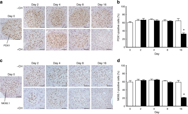
Fig. 7.
Immunohistochemistry and quantification of PDX1 (a, b) and NKX6.1 (c, d) in pancreatic sections of NZO mice after a diet change from carbohydrate-free (−CH) (white bars) to carbohydrate-containing diet (+CH) (black bars). (a, c) Sections embedded in paraffin at the indicated day were haematoxylin-stained and immunostained with the respective antiserum and developed with DAB as described in Methods. Scale bars, 50 μm. (b, d) Quantifications of nuclei positive for PDX1 (b) and NKX6.1 (d) were performed for three to six mice per group; *p ≤ 0.05 for –CH vs +CH at the indicated time points

