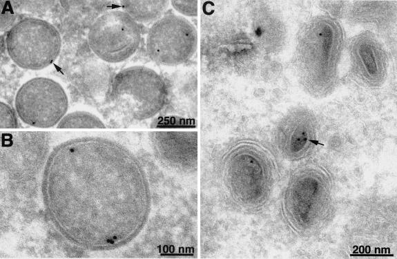
FIG. 10.
Localization of F10V5 by immunoelectron microscopy. BS-C-1 cells were infected with vF10V5i at a multiplicity of infection of 10 in the presence of 50 μM IPTG. Twenty-two hours after infection, the cells were fixed in paraformaldehyde, cryosectioned, and incubated with anti-V5 MAb followed by rabbit anti-mouse IgG and protein A conjugated to colloidal gold. Arrows point to representative gold grains. Electron micrographs are shown with scale bars.

