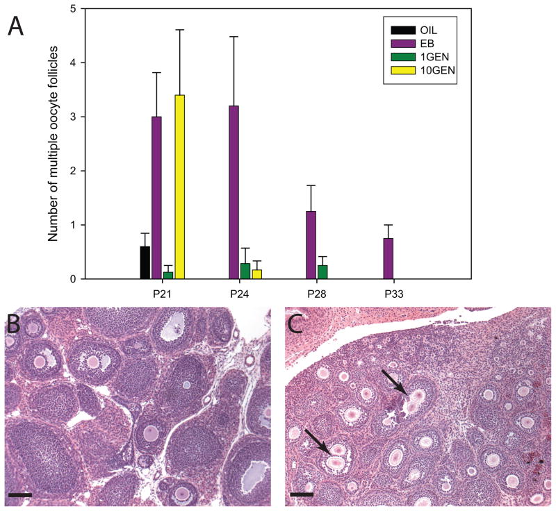
Fig. 3.
(A) The average number of multiple oocyte follicles (MOFs) found in the different exposure groups at each age, with control (OIL) animals having no MOFs by P24, EB exposed animals having MOFs at all time points and 10GEN exposed animals having MOFs through P24. Because of technical issues, tissue from P28 and P33 10GEN females, and P33 OIL animals was not available. Representative images (taken at 20x) depicting ovarian follicles from P21 OIL (B) and 10GEN (C) females. MOFs (arrows) were readily apparent in the 10GEN ovary. (scalebar = 40μm)

