Fig. 1.
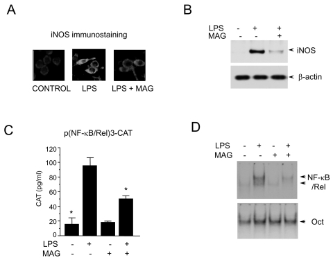
Inhibition of macrophage activation by magnolol. (A) RAW 264.7 cells (5×10⁵ cells/ml) incubated with magnolol (50 µM) in the presence of LPS (200 ng/ml) for 24 hr on cover slide in 12 well plates. Cells were subjected to immunohistochemical staining using an antibody specific for murine iNOS. Immunoreactivity of iNOS was localized along the margins of the cytoplasm in control group. (B) Cells were treated with magnolol in the presence of LPS (200 ng/ml) for 24 hr. Cell lysates were then prepared and subjected to Western immunoblotting. (C) RAW 264.7 cells were transfected with p(NF-κB/Rel)3-CAT by DEAE dextran method. Twenty-four hours after transfection, cells were treated with the magnolol in the presence or absence of LPS (200 ng/ml) for 18 hr. Cell extracts were then prepared and analyzed for the expression of CAT using CAT ELISA kit. (D) Cells (5×10⁵ cells/ml) were incubated with magnolol (50 µM) in the presence or absence of LPS (200 ng/ml) for 2 hr. Nuclear extracts (5 µg/ml) were then isolated and analyzed for the activity of NF-κB/Rel and Oct.

