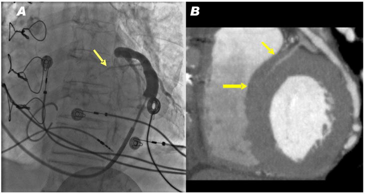
Figure 9.
Representative human venograms. (A) A pressurized venogram in a patient undergoing cardiac resynchronization therapy. A basal septal perforator vein was evident (arrow) in all 8 patients with evaluable angiograms. (B) A CT angiogram showing a basal septal perforator vein (arrows) apparently suitable for cerclage.

