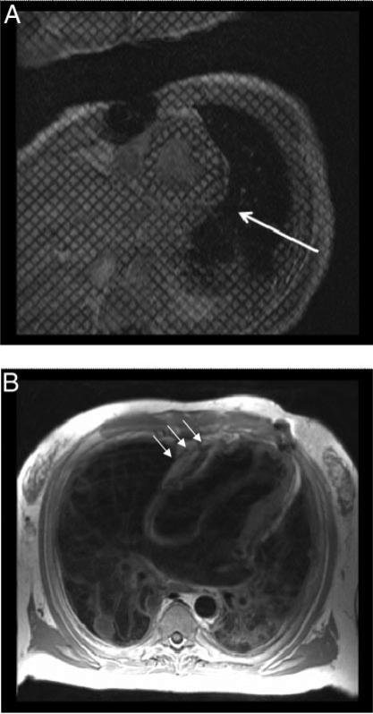
Figure 13.
Cardiovascular magnetic resonance findings associated with pericardial disease. Panel A: a short-axis, cine-tagged imaging is provided. Along the posterior wall of the left ventricle (white arrow), tag deformation is absent, indicating pericardial adhesions. Panel B: dark blood T1-weighted spin echo images are provided, indicating thickened pericardium along the anterior surface of the right ventricle and corresponding tubular deformity of the ventricles. Advanced lung disease is also noted.

